|
The following case studies are examples of the uses of diagnostic digital infrared imaging in clinical practice.
Please choose from the following sections:
BREAST THERMOGRAPHY
Case #1 -
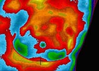
Left Breast TH 4 – Abnormal
Initial Infrared Image
|
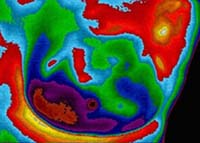
Left Breast TH 2 – Normal Vascular
Post-lumpectomy Image
|
The above images are of the left breast of a 51 year old patient presenting with a non-tender mass in the 6 o'clock position along the inferior margin of the breast (black cross-hairs on image). The patient's gynecologist discovered the lump on physical examination and described it as an indurated mass the size of a kidney bean. Follow up mammographic and ultrasound studies were performed with similar findings in size and an interpretation of mild architectural change of no concern. The above left image (Initial infrared examination – TH 4) shows significant signs of angiogenesis along with a focal signal increase over the site of the mass (black cross-hairs on image). The patient followed up with a surgical consult and elected to have a lumpectomy. Her surgery revealed a large ovoid mass extending back toward the chest wall. Cytological examination found the cells to be pre-cancerous. The patient's surgeon informed her that she was very lucky to have opted for a lumpectomy, as the size of the mass alone, when it turned to cancer, would have been very serious. The above right image (Post-lumpectomy – TH 2) was taken one month following resection of the mass and shows complete resolution of the angiogenic activity along with an isothermic signal over the site of the resected mass.
|